SENJIE Healthy & Technology SENJIE Healthy & Technology

What Is the Best Neurosurgery Equipment in 2026?

Time:2026-09-26 Author:Isabella
0%

Choosing the best Neurosurgery Equipment in 2026 is not about finding one machine that suits every operating room. It means matching dependable tools to the procedure, clinical team, and hospital resources. The stakes are substantial: a 2024 analysis of the Global Burden of Disease Study 2021, published in The Lancet Neurology, estimated that 3.4 billion people experienced neurological conditions worldwide. That figure describes a broad health burden, not demand for any particular device.

Precision matters. A surgical microscope can support fine work around delicate vessels, while neuronavigation helps teams relate instruments to preoperative images. Intraoperative imaging and neurophysiological monitoring may add useful information during selected procedures. Yet each tool has limits. Registration can drift, images can become outdated as tissue shifts, and monitoring signals require skilled interpretation. More technology is not automatically better.

A practical 2026 comparison should weigh clinical evidence, image quality, usability, training, maintenance, and total cost—not promotional claims alone. For hospitals, uptime matters as much as a feature list; a sophisticated system is less useful when specialist support or replacement parts are unavailable. There is no universal winner. The right choice depends on the operation, staff expertise, and local workflow, and that answer may change between centers. This guide examines leading equipment categories and the questions buyers and clinicians should ask before adopting them. Some trade-offs remain uncomfortable, and evidence does not resolve every purchasing decision.
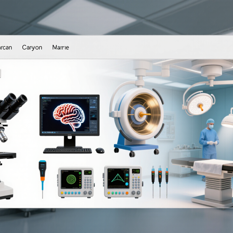
What Is the Best Neurosurgery Equipment in 2026?

How “Best” Neurosurgery Equipment Is Evaluated in 2026

What Is the Best Neurosurgery Equipment in 2026?

How “Best” Neurosurgery Equipment Is Evaluated in 2026

In 2026, “best” means clinically suitable, dependable, and supportable—not simply newest. Evaluation should test image clarity, navigation accuracy, instrument handling, setup time, sterilization compatibility, and performance in realistic workflows. Ask surgeons, nurses, biomedical engineers, and procurement staff to assess the same system. Record failures, calibration drift, and service response times. Small details matter: Can staff read the display under operating-room lights? Does a cable cross the assistant’s path?

Evidence should extend beyond specifications. In an eight-hospital study published in the New England Journal of Medicine, complications fell from 11.0% to 7.0% after surgical checklist implementation; deaths fell from 1.5% to 0.8% (Haynes et al., 2009). This was not an equipment trial. It does, however, show why tools must support safe team workflows. ECRI’s 2025 Top 10 Health Technology Hazards report also highlights risks involving connected devices and technology management. Buyers should review cybersecurity support, software updates, training needs, and lifecycle costs—not just purchase price. Evidence has limits; local workflow testing still matters.

Tips: Simulate a case before purchase. Time setup, test backup power, and ask staff to identify one failure point. Document what changed.

Core Imaging and Navigation Systems for Neurosurgical Procedures

In 2026, the best neurosurgery equipment is not a single device. It is an imaging and navigation setup that fits the procedure, anatomy, and operating-room workflow. Preoperative MRI helps define soft-tissue boundaries, while CT can clarify bone and implanted hardware. During surgery, updated images may help reveal changes that earlier scans cannot show. Precision matters.

Navigation systems link these images to the patient’s position, giving surgeons a moving reference on screen. Accurate registration is essential: even a small mismatch can make a displayed target less trustworthy. Teams should check alignment after positioning and revisit it when anatomy shifts. No system removes uncertainty. A screen is a guide, not the patient.

For a practical comparison, assess image quality, update speed, instrument tracking, and how easily staff can verify calibration. Intraoperative imaging can be valuable when visibility changes during a case, but it adds space, setup time, and workflow demands. Test the full setup in realistic conditions, including draping and staff movement. A useful detail is whether key images remain readable from the surgeon’s working position. I would also ask how often the system needs recalibration; that detail is easy to overlook. The right choice supports careful judgment rather than promising perfect accuracy.

What Is the Best Neurosurgery Equipment in 2026? - Core Imaging and Navigation Systems for Neurosurgical Procedures
System Primary Role Common Neurosurgical Uses Key Strengths Important Limitations Best Fit
Preoperative MRI Creates detailed images of the brain, spinal cord, and soft tissues before surgery. Brain tumor assessment, vascular and inflammatory lesion evaluation, and planning of many cranial procedures. Excellent soft-tissue contrast; does not use ionizing radiation; multiple sequences can help characterize anatomy and pathology. Images are acquired before surgery and may not reflect tissue movement during an operation. Some implants or patient conditions require additional safety assessment. Preoperative soft-tissue assessment and surgical planning.
CT and CT angiography Provides cross-sectional imaging of bone, acute blood, and—when contrast is used—vascular structures. Trauma, skull and spine anatomy, hemorrhage assessment, and selected vascular or bony procedures. Fast acquisition; strong depiction of bone; widely used in urgent imaging and image-guided planning. Uses ionizing radiation. Soft-tissue contrast is generally lower than MRI, and contrast-enhanced studies require consideration of contrast-related risks. Urgent evaluation, bone-focused planning, and cases where rapid imaging is important.
Intraoperative MRI Obtains MRI images during surgery to reassess anatomy and residual target tissue. Selected brain tumor resections and procedures where updated soft-tissue imaging may affect the next surgical step. Can show changes that occur after preoperative imaging and help assess the resection before the operation is completed. Requires specialized facilities, MRI-compatible instruments, and carefully managed workflow. Imaging and room transitions can add time; it is not appropriate for every case. Selected procedures where updated MRI findings could change intraoperative decisions.
Intraoperative CT Provides updated cross-sectional images in or near the operating room. Spinal instrumentation, selected cranial procedures, and assessment of bone, hardware position, or acute changes. Useful for checking anatomy and implant position during a procedure; CT depicts bone well and can be acquired without moving the patient to a separate imaging department in some setups. Uses ionizing radiation. Image quality and workflow depend on the scanner configuration, patient positioning, and operating-room setup. Procedures where intraoperative confirmation of bony anatomy or hardware position is valuable.
Image-guided neuronavigation Displays instrument location in relation to registered preoperative CT or MRI images. Cranial tumor surgery, biopsy, ventricular access, and selected skull-base or spinal procedures. Supports orientation to patient-specific anatomy and can help plan trajectories while reducing reliance on surface landmarks alone. Registration and tracking errors can affect displayed accuracy. Brain shift during cranial surgery can make preoperative images less representative of current anatomy. Procedures requiring image-based localization or trajectory planning, used alongside direct clinical judgment.
Intraoperative ultrasound Provides real-time imaging through a probe during surgery. Selected brain tumor resections, cyst or fluid-space assessment, and procedures where updated intraoperative views are useful. Can provide repeat imaging during the operation without ionizing radiation; may help visualize anatomy after surgical exposure. Image quality and interpretation depend on the acoustic window, probe handling, and operator experience. Images may be less intuitive than MRI for some users. Cases where real-time, repeatable imaging is useful and an adequate acoustic window is available.
Stereotactic frame or frameless guidance Establishes a coordinate-based or image-registered pathway to a target. Brain biopsy, functional procedures, and other interventions requiring precise target localization. Provides structured target and trajectory planning; frame-based methods use a rigid reference, while frameless methods can support a less restrictive setup. Frame-based methods require frame placement and can be uncomfortable or time-consuming. Frameless guidance depends on reliable registration and may be affected by tracking or anatomical changes. Targeted procedures where stereotactic localization is part of the planned technique.

Selection note: No single system is best for every neurosurgical procedure. Equipment choice depends on the clinical objective, patient-specific anatomy, available expertise, workflow, and institutional safety protocols.

Essential Instruments for Cranial and Spinal Surgery

For cranial surgery, the essential setup is not a single “best” device, but a dependable system. A guarded high-speed drill, fine bipolar forceps, microsurgical instruments, controlled suction, and an operating microscope support precise work around delicate tissue. Navigation and neurophysiological monitoring can add useful information, but they do not replace anatomical judgment. Keep the instrument tray focused. Too many tools can slow a handoff.

Spinal procedures need a different balance: Kerrison-style punches, curettes, nerve-root retractors, pedicle probes, and suitable fixation instruments. Intraoperative imaging helps confirm levels and hardware position; neuromonitoring may help detect changes during selected procedures. Fit matters. A poorly matched instrument can obstruct access or reduce control, even when it is technically advanced.

Reliable support matters as much as the instrument list. Check sterilization compatibility, calibration, maintenance records, and staff familiarity before use. The Lancet Commission on Global Surgery reported in 2015 that about 5 billion people lacked access to safe, affordable surgical and anesthesia care, with an estimated 143 million additional procedures needed each year. These figures describe access, not neurosurgical equipment performance. Still, they underline why durable, serviceable equipment and practical training deserve attention. No checklist is perfect; local anatomy, procedure, and team experience should guide final selection.

Robotic, Monitoring, and Visualization Technologies

In 2026, neurosurgical equipment is strongest when robotics, monitoring, and visualization work as one system. Robotic guidance can help surgeons plan trajectories and place instruments with precision, but registration errors and shifting anatomy still matter. A robot is not an autopilot. Evaluate accuracy, setup time, and how reliably the team can respond when the plan changes.

Visualization can influence decisions in the operating room. In a 2011 randomized trial published in The Lancet Oncology, intraoperative MRI helped achieve complete tumor resection in 96% of patients, compared with 68% using conventional surgery. That result applies to the studied patients and setting; it is not a guarantee for every tumor. Clear microscope views, image-guided navigation, and updated scans can help surgeons see boundaries, though images may still differ from what the tissue reveals.

Monitoring adds another layer. The American Academy of Neurology’s evidence review on intraoperative spinal monitoring found that significant signal changes can help predict neurological injury. Teams should agree on alert thresholds and response steps before surgery. A quiet monitor is not proof of safety. Nor does every alert mean damage. The human interpretation remains essential, and that part can be imperfect.

How Hospitals Select Equipment for Different Neurosurgical Needs

In 2026, the best neurosurgery equipment is the system that fits a hospital’s cases, staff, and operating rooms—not simply the newest system. A cranial tumor service may prioritize an operating microscope, neuronavigation, and reliable intraoperative imaging. A spine service may need compatible imaging, surgical drills, and neuromonitoring. Fit matters. Before purchase, surgeons, nurses, biomedical engineers, and infection-prevention staff should assess room space, setup time, sterile processing, training, and service support.

The Lancet Commission on Global Surgery estimated that five billion people lacked access to safe, affordable surgical and anesthesia care, while 143 million additional procedures were needed each year in low- and middle-income countries. These figures do not specify neurosurgical equipment needs, but they underline why purchasing decisions should consider access, upkeep, and actual clinical demand. A practical review can compare case volumes, equipment downtime, staff competency, and the cost of recurring accessories. A high-spec system is a poor choice if it sits unused or cannot be maintained.

Hospitals should also test how devices work together at the bedside and in the operating room. For example, teams can rehearse a navigation setup, check image transfer, and confirm that cables leave clear space around the table. ECRI’s Top 10 Health Technology Hazards for 2024 highlights how technology problems can disrupt patient care, supporting careful review of connected-device security and downtime plans. Downtime hurts. No checklist removes uncertainty; even experienced teams may overlook workflow friction until real cases begin.

FAQS

What makes neurosurgery equipment “best” in 2026?

It should fit the procedure, work reliably, and receive dependable support. Newness alone is not enough.

How should hospitals evaluate equipment before buying it?

Have surgeons, nurses, engineers, and purchasing staff test the same setup. Time installation, check display visibility, and record calibration problems. Simulate a case.

What should teams check in imaging and navigation systems?

Compare image clarity, update speed, instrument tracking, and calibration checks. Recheck alignment after positioning or when anatomy shifts. A screen is only a guide.

When can intraoperative imaging be useful?

Updated scans may help when visibility changes during surgery. They also require space, setup time, and staff coordination. Test them with drapes in place.

Can robotic guidance guarantee accurate instrument placement?

No. Registration errors and shifting anatomy can affect guidance. Assess setup time and how easily staff can adjust when plans change. Not autopilot.

What can monitoring signals tell surgical teams?

Significant signal changes may help predict neurological injury. Teams should agree on alert thresholds and response steps before surgery. A quiet monitor is not proof of safety.

Do study results prove that a system will work for every patient?

No. One trial reported complete tumor resection in 96% of patients with intraoperative imaging, versus 68% with conventional surgery. Those results apply to its studied setting, not every case.

What safety and cost factors should buyers consider?

Review cybersecurity support, software updates, staff training, service response, and lifecycle costs. A checklist study found fewer complications after implementation, but it did not test equipment. Local testing still matters.

Conclusion

In 2026, the best Neurosurgery Equipment is not defined by a single device but by how well a complete set of tools supports safe, precise, and efficient care. Evaluation may consider imaging quality, navigation accuracy, ease of use, reliability, compatibility with existing systems, and the needs of both patients and clinical teams. Imaging and navigation systems help surgeons understand anatomy and plan procedures, while specialized instruments support a range of cranial and spinal operations.

Robotic assistance, physiological monitoring, and advanced visualization can further support surgical planning and decision-making. However, the most suitable equipment depends on each hospital’s procedures, resources, staff expertise, and patient population. A thoughtful selection process weighs clinical value, training requirements, maintenance, and long-term costs rather than focusing on technology alone. By matching these factors to local needs, hospitals can build a practical equipment environment that supports consistent, patient-centered neurosurgical care.

Isabella

Isabella

Isabella is a dedicated marketing professional with a sharp focus on driving brand growth and engagement through strategic content creation. With an extensive background in digital marketing, she combines her passion for storytelling with her keen understanding of industry trends to deliver......